Tuesday, 31 July 2012

Diffusion

Learning Outcomes

  • Define the process of diffusion as the net movement of molecules from a region of high concentration to a region of low concentration.
  • Define the process of osmosis as the net movement of water molecules from a region of higher water potential to a region of lower water potential across a partially permeable membrane.
  • Explain that osmosis is a subset of diffusion, and that osmosis is restricted to the net movement of water molecules, whilst diffusion involves the movement of any type of molecules. (Note: a partially permeable membrane does not necessarily have to be present for diffusion.)
  • Explain how animal and plant cells behave differently in solutions of varying water potential.
  • Appreciate the importance of diffusion and osmosis to living systems, and list examples of both processes.

Diffusion is the net movement of particles from a region of higher concentration to a region of concentration.
                         
Concentration =  amount of  a substance           --> amount -> mass (g/kg)
                           volume of a fluid (water)        volume -> cm3/ml

High concentration--> amount of substance UP
                                    volume of fluid DOWN

Low concentration--> amount of substance DOWN
                                    volume of fluid UP

WRONG example: Mom is cooking in the kitchen and you can smell the food ( NOT diffusion)

1. Is diffusion a spontaneous process (no energy required)?
 
    No, diffusion does not need energy to occur.

  •  Substances tend to spread from an area where they are more concentrate to an area where they are less concentrated.
  • Two or more substances can become evenly distributed (reach equilibrium) even without external interventions  


2. Why does diffusion not occur instantaneously in the living world?

    There is air in the living world, slowing down the rate of diffusion.

3. What are the factors affecting the diffusion rate?

  • Temperature
  • Presence of air 
  • Size

Concentration Gradient

Concentration
  • A measure of the amount of a substance in a specific volume 
Concentration Gradient
  • The concentrating gradient between points A and B is the difference in concentration between points A and B.
Sugar molecules diffuses from point A to point B
We can say: Sugar molecules diffuses down the concentration gradient from point A to B.

  • Particles diffuses down the concentration gradient
  • The larger the concentration gradient, the faster the rate of diffusion.

Two types of membranes:
  • Permeable membrane - Allow all substances to pass through (much large gates/holes)
  • Partially permeable membrane - Allows some substances to pass through. (small gate/holes)

Application of diffusion in biology

The visking tubing encloses a solution of starch while the beaker contains iodine solution. Starch reacts with iodine to form a dark blue complex. Only the starch in the visking tubing will turn blue. 
What type of membrane is the visking tubing?
Explain.

partially permeable. Only the iodine managed to pass through the visking tubing. Starch, which has big particles, could not pass through the visking tubing.


Conclusion 

  • Diffusion is an important process where substances are moved without use of energy.
  • It is the net movement of particles (or molecules; or ions) from a region of higher concentration to a region of lower concentration.
  • Thus the movement is down a concentration gradient.
  • It is important to bear in mind that
          -The movement is random
          -The greater the concentration gradient, the faster the rate of diffusion.

Monday, 30 July 2012

Biomes

Biomes
A major ecological community of organisms adapted to a particular climatic or environmental condition on a large geographic area in which they occur.  
Biomes may be classified into:

  1. Terrestrial biomes or land biomes - e.g. tundra, taiga, grasslands, savannas, deserts, tropical forests, etc. 
  2. Freshwater biomes - e.g. large lakes, polar freshwaters, tropical coastal rivers, river deltas, etc. 
  3. Marine biomes - e.g. continental shelf, tropical coral, kelp forest, benthic zone, pelagic zone, etc. 
 Word origin: bi: variant of bio-, esp. before a vowel + -ome: New Latin -ōma, -ōmat.

PT
Tundra Biome 
Located at latitudes 55° to 70° North, the tundra is a vast and treeless land which covers about 20% of the Earth's surface, circumnavigating the North pole. It is usually very cold, and the land is pretty stark. Almost all tundras are located in the Northern Hemisphere. Small tundra-likeareas do exist in Antarctica in the Southern Hemisphere, but because it is much colder than the Arctic, the ground is always covered with snow and ice. Conditions are not right for a true tundra to form. Average annual temperatures are -70°F (-56°C).

Ecology

Factors
oxygen salinity light
pH
-may be influenced by photosynthetic activity of aquatic plants

The more acidic a substance, the sourer it is. 
The higher the acidic, the lower the pH
7pH is neutral. 

acids --> pH1-6.9

alkali pH --> 7.1-14

Water
(neutral) --> pH7.0

Biotic Factors

Symbiosis
1.Mutualism +/+ positive! (Hermit crab)
2. Commensalism (one benefits;the other unaffected) +/0
3. Parasitism (parasite benefits and host harmed) +/- dog and fleas. worms in human body

1. competition -/- (Interspecific- a form of symbiosis) (Intraspecific-same species)
2. Predation (one feeds on the other) +/-

Producer-Primary consumer->secondary consumer->tertiary consumer-> quaternary consumer

Saturday, 28 July 2012

Lab Practical 4

We had to scrub our cheek cells with a toothpick, rub it on a glass slide and place it under a microscope! After that, we placed a tiny leaf of a hydrilla plant under the microscope and here are the pictures(: It wasn't easy to take photographs, putting the camera in front of the eyepiece:0

Leaf of a hydrilla plant

Leaf of a hydrilla plant

Lab Practical 3

Lab Practical 3. First lesson learning how to use a light microscope.
Our class learnt the different types of microscopes: light microscopes(the one we used), Electron Microscope and more! We learnt how to calculate the magnification (eyepiece 10X multiplied by various objective lenses) and there were three objective lenses: scanning objective lens, low objective lens and high-power objective lens.(ascending order) Next, we learnt how to prepare a slide(which was to be put on the stage of the microscope later on).

Here are the steps:
1. Wash the glass slide thoroughly under running water. (You don't want to see thumbprints on the glass slide under the microscope!!) **You must must hold it by its sides, not the wide glass surface**

2. After you have dried all the water off the glass slide, put the substance you want to observe under the microscope.

3. For me, it was newspaper. So I had to put some water because newspaper is a solid substance. (If it is not coloured, [like cheek cells] you will have to put a drop of iodine solution or a colored liquid!)
(If it is in liquid form already, you do not need to put any any thing.)

4. Next, carefully slide the cover slip down onto the glass slide! (you don't want to see any huge air bubbles under the microscope! You will see a black line) (I had experienced this before; I saw some black dots under the microscope and mistook it for the substance.__.) To prevent any air bubbles, you will have to use a sharp object (I have been using toothpicks) to drag the cover slip along the glass slide. 

5. We have finally prepared the slide!:D

So after so many adjustments of the stage....

I came up with this wonderfully huge picture of the newspaper and the three fibered strings! (the newspaper was [] <--this size)

A tiny piece of newspaper with the letter 'S' under a microscope
The letter 'S' is upside-down and laterally inverted!

3 different coloured strings under a light microscope.
If you focus on one string, the other strings will look blur!

Drawing of Human Cheek Cells and Plant Cells

Title: Drawing of Human Cheek Cells 

Title: Drawing of Hydrilla Leaf Cells 

Complex Drawing of an Animal Cell

A Complex Drawing of an Animal Cell

Thursday, 26 July 2012

Biology Discussion - Cells

Red Blood Cell

Structure and functions

  • Function: The red blood cell delivers oxygen to body tissues via blood flow through the circulation. They take up oxygen in the lungs or gills and release it while squeezing through the  body’s capillaries.

  • Structure:
  • Biconcave discs, having a depressed center on both sides. (These depressed centers allow the cells to have more cell membrane surface we tend to use the phrase “higher surface area to volume ratio” which can be exposed to diffusing oxygen while transiting the lungs. This structure also allows them to be more flexible when negotiating tight passages.)
  • 7.8 micrometers in diameter

  • Does not have nucleus and most organelles such as mitochondria to accommodate maximum space for haemoglobin.(the compound that carries oxygen through the body.)
  • check this website: http://www.wisc-online.com/objects/ViewObject.aspx?ID=ap14604
  • Red blood cells are red only because they contain a (protein chemical ← wrong term.  Haemoglobin is a protein, we don’t call it a protein chemical) called hemoglobin which is bright red in colour
  • The main function of the red blood cell is to transport oxygen from the lungs, to the other tissues and cells of the body. The other function of the red blood cell is to partly carry carbon dioxide, which is a
    • Does not have nucleus and most organelles such as mitochondria to accommodate maximum space for haemoglobin.(the compound that carries oxygen through the body.)
    • check this website: http://www.wisc-online.com/objects/ViewObject.aspx?ID=ap14604
    • Red blood cells are red only because they contain a (protein chemical ← wrong term.  Haemoglobin is a protein, we don’t call it a protein chemical) called hemoglobin which is bright red in colour
    • The main function of the red blood cell is to transport oxygen from the lungs, to the other tissues and cells of the body. The other function of the red blood cell is to partly carry carbon dioxide, which is a
      • Does not have nucleus and most organelles such as mitochondria to accommodate maximum space for haemoglobin.(the compound that carries oxygen through the body.)
      • check this website: http://www.wisc-online.com/objects/ViewObject.aspx?ID=ap14604
      • Red blood cells are red only because they contain a (protein chemical ← wrong term.  Haemoglobin is a protein, we don’t call it a protein chemical) called hemoglobin which is bright red in colour
      • The main function of the red blood cell is to transport oxygen from the lungs, to the other tissues and cells of the body. The other function of the red blood cell is to partly carry carbon dioxide, which is a
        • Does not have nucleus and most organelles such as mitochondria to accommodate maximum space for haemoglobin.(the compound that carries oxygen through the body.)
        • check this website: http://www.wisc-online.com/objects/ViewObject.aspx?ID=ap14604
        • Red blood cells are red only because they contain a (protein chemical ← wrong term.  Haemoglobin is a protein, we don’t call it a protein chemical) called hemoglobin which is bright red in colour
        • The main function of the red blood cell is to transport oxygen from the lungs, to the other tissues and cells of the body. The other function of the red blood cell is to partly carry carbon dioxide, which is a waste product of metabolic activities in the body.


        Xylem Vessels/ Xylem Cells


        Consist of dead hollow cells because the walls are lignified and the cell contents disintegrate. The lignin makes the cell wall impermeable so they are in effect waterproof. It also makes the vessels extremely strong and prevents them from collapsing. They have a wide lumen and are linked end to end to create a long, hollow tube since the end cell walls have one or many perforations in them. This allows the transport of large volumes of water. The sidewalls have bordered pits (unlignified areas) to allow lateral movement of water. Xylem vessels are found in angiosperms


        Intestinal Cell (sometimes known as intestinal epithelial)


        Location: the small intestines

        Structure and characteristics:

        - Contain many membrane-bound vacuoles
        - Aglycocalyx surface coat contains digestive enzymes. ← no need to know this for now, it is beyond your syllabus
        - Microvilli on the apical surface increase surface area ← once again, the portion that is underlined should be re-written as “increase surface area to volume ratio” for the digestion and transport of molecules from the intestinal lumen

        The points stated below refers to the functions of the intestine, which are facilitated by the intestinal epithelial cells
        • Ion uptake
        • Water uptake
        • Sugar uptake
        • The main function of intestinal cells is associated with secreting
                • digestive juices into the lumen (the inner cavity of an intestine or blood vessel).


        Root hair Cell


        Structure + Function

        1. Elongated structure that protrudes out to the soil
        - This is to increase the surface area to volume ratio; thereby increasing the rate of uptake of water from the soil to the cell.

        2. Large vacuole

        - The root hair cell has a large central vacuole to maximize the amount of water capacity of the cell; thus, the cell is able to absorb and store more water.

        3. Cell sap

        - The cell sap of the root hair cell has a lower water potential than the water in the soil ← Good. We will learn what is water potential in the later lessons after ecology. Thus, the water from the soil moves into the cell via osmosis.

        Read more: http://wiki.answers.com/Q/What_is_the_structure_of_root_hair_cells#ixzz21S1j6XeB

        For the points below, it is more of how the various structures in the cell contributes to the function of a cell.  May be said for plant cells in general, and is not specific to only the root hair cell.

        4. Nucleus

        -Contains contains chromatin material, consisting of the DNA if the cell which is important (inherited by the daughter cells)

        5. Plasma membrane

        -Controls the movement of substance into and out of the cell and is used for cell
            • identification.

        6. Cell Wall

        -The cell wall is a strong surface, surrounding the plasma membrane, which protects the cell and give it its shape. It also prevents expansion when too much water enter the cell.

        7. Cytosol

        -The cytosol is made up of water, salts and organic molecules and many enzymes that speed up reactions. It is important as it suspends the cell organelles within it.

        Read more: http://sst-health-science-class-107.blogspot.sg/2010/01/root-hair-cell.html

Monday, 23 July 2012

Differences between Animal and Plant Cells

Plant cells have a  large central vacuole while animal cells have many small vacuoles.
Plant cells have a cell wall while animal cells does not have a cell wall.
Plant cells have a fixed shape while animal cells have a irregular shape.
Plant cells have chloroplast while animal cells does not.

Monday, 16 July 2012

Cell Structure and Function

Cell Structure and Function

Cell theory

  • All living things are made up of cells.
  • Cells are the smallest working units of all living things.
  • All cells come from pre-existing cells through cell division.

Definition of cell

A cell is the smallest unit that is capable of performing life functions.

Examples
  • Amoeba
  • Plant stem
  • bacteria
Two types of cells 
  • Prokaryotic
  • Eukaryotic
Prokaryotic
  • Do not have organelles (specialised structures in cells) surrounded by membranes
  • Few internal structures
  • one-celled organisms e.g. bacteria

Eukaryotic
  • Contain organelles surrounded by membranes 
  • Most living organisms e.g. plant, animal, fungi

Plant
Animal
Cell Wall
  • Most commonly found in plant cells&bacteria
  • Surrounds the cell membrane
  • Rigid structure that maintains the shape, supports & protects cells
  • Permeable to small molecules and small proteins only 
Membrane
  • Selectively/partial* permeable membrane of cell that controls movement in and out of the cell.
*XXXXX semi- permeable XXXXX
Nucleus
  • Contain chromatins that control cell activities 
  • Chromatin contain DNA which is the genetic material
  • DNA contain instructions for traits & characteristics and to carry out the cell's function
  • Separated from cytoplasm by nuclear membrane 
**Chromatin is a combination of DNA and protein. DNA winds around the protein

Cytoplasm
  • Gel-like mixture
  • Surrounded by cell membrane
  • Contains organelles
Mitochondria
  • Referred to as the  "powerhouse" of the cell.
  • The food we eat is transformed into energy (ATP) for the cell and our bodies.
Endoplasmic Reticulum
  • An interconnected network of tubs and vesicles
  • Synthesis of(Make) proteins, fats, steroid
  • Transports materials around in cell
  • Smooth type: lacks ribosomes
  • Rough type (in picture): ribosomes e bedded in surface
Ribosomes 
  • Each cell contains thousands
  • Make proteins
  • Found on endoplasmic reticulum & floating throughout the cell 
  • Why on endoplasmic reticulum? Once the ribosomes make the protein, the protein will go into the endoplasmic reticulum. Endoplasmic reticulum folds the proteins into the correct shape so that the proteins can function properly.

Golgi Apparatus(whole stack)/Golgi Bodies(smaller) 
  • Works closely with the ER Primary function is to process and package complex molecules such as proteins and fats that are made by cell
  • Brings these products to the surface of the cell where they can be secreted
  • Other secretions include hormones, antibodies and enzymes.
Lysosome
  • Contain digestive enzymes
  • Digest excess or worn-out organelles, food particles and engulf bacteria or viruses
  • Also help repair worn-out plasma membrane
  • They also provide sugars, amino acids and bases which are the foundations of macromolecules
  • Cell breaks down if lysosome explodes

Vacuoles


  • Membrane-bound sacs for storage, digestion and waste removal
  • Central large vacuole-help plant cells maintain shape
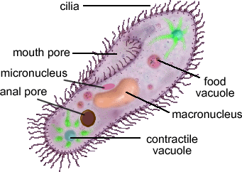
  • Food vacuole: formed by phagocytosis (when the cell tries to engulf something.


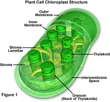
Chloroplast
  • Usually found in plant cells (some bacterias have chloroplast too)
  • Contains green chlorophyll
  • Where photosynthesis takes place
  • Converts light energy into chemical energy in glucose.


Monday, 9 July 2012

Classification

Why do we need to classify organisms?

- easier to find organisms
- easier to see the similarities and differences of the organisms
- easier to see trends in the organisms
- easier to retrieve information from the classification


How many species are there?

:13 billion

This is only 5% of all organisms that ever lived!

What is classification?


  • Classification is the arrangement of organisms into orderly groups based on their similarities
  • Classification is also known as taxonomy
  • taxonomists are scientists that identify & name organisms                                                                    

Benefits of classifying

  • Accurately & uniformly names organisms
  • Prevents misnomers such as starfish and jellyfish that aren't really fish
  • Use same language (Latin or some Greek) for all names. 


Binomial nomenclature
Genus      species

Problems in classifying organisms

  • Species are always changing.
  • Mutation
  • Interbreeding
Naming of Organisms


Print form

Homo sapiens

Written form

Homo sapiens


Classification Group

  • Taxon (Taxa-plural) is a category into which related organisms are placed.
  • There is a hierarchy of groups(taxa) from broadest to most specific
  • Domain-Kingdom-Phylum-Class-Order-Family-Genus-Species

Domains
  • Broadest, most inclusive taxon
  • three domains
  • Archaea and Eubacteria are unicellular prokaryotes(no nucleus or membrane-bound organelles)
  • Eukarya are most complex and have a nucleus and membrane-bound organelles
Taxons
  • Most genera contain a number of similar species, with the exception of Homo that only contains more different types of organisms than the preceding category.
Examples

Human classification


Domain Eukarya-->cells with nuclei
King Animalia--> Multicellular, motile, ingestion of food
Phylum Chordata--> Dorsal supporting rod and nerve cord
Class Mammalia--> Hari, mammary glands
Order Primates--> Adapted tp climb trees
Fmaily Hominidae--> Adpted to walk erect
Genus Homo...
Species


Pictures taken from http://sarahfoo26106biologyjournal.blogspot.sg/